
The dynamic assembly, remodeling, and turnover of actin networks drives cellular processes
ranging from cell motility, endocytosis, and phagocytosis to cell division, cell and tissue
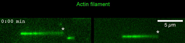
morphogenesis, and neuronal pathfinding. Here, we describe a new actin regulatory activity that changes understanding of how actin networks can be turned over. In a collaborative project with Bruce Goode’s lab, postdocs Shashank Shekhar and Greg Hoeprich used microfluidics-assisted total internal reflection fluorescence (TIRF) microscopy to show that mammalian twinfilin, an evolutionary conserved ADF/cofilin-homology protein, accelerates depolymerization at newly-assembled (ADP-Pi) but not older (ADP) actin filaments, even under assembly-promoting conditions (i.e., at G-actin concentrations above the critical concentration). Our data suggest that twinfilin molecules interact processively with the barbed end of the filament as it shrinks, blocking ATP-actin subunit addition while allowing ADP-Pi subunit dissociation. These novel activities of twinfilin reveal that cells have machinery that can bypass the normal filament aging process and induce the depolymerization of barbed ends as needed. These results may explain known genetic interactions between twinfilin and cofilin, and localization of twinfilin to the tips of filopodia and stereocilia, where actin filament barbed ends are clustered.
[ensemblevideo contentid=94fc0a2d-eaad-4ccb-84ac-7ebf799ba3ec autoplay=true]
10.1083/jcb.202006022Shekhar S, et al., Twinfilin bypasses assembly conditions and actin filament aging to drive barbed end depolymerization.
Journal of Cell Biology 220, e202006022 (2021)
 The new study shows that Cofilin and one other protein (Srv2/CAP) intimately collaborate at one end of the actin filament to accelerate subunit dissociation by over 300-fold! These are the fastest rates of actin depolymerization ever observed. Further, these results establish a new paradigm in which a protein that decorates filament sides (Cofilin) works in concert with a protein that binds to filament ends (Srv2/CAP) to produce an activity that is orders of magnitude stronger than the that of either protein alone.”