Tag Archives: NIGMS-funded

Regulation of replication origin licensing by ORC phosphorylation reveals a two-step mechanism for Mcm2-7 ring closing.

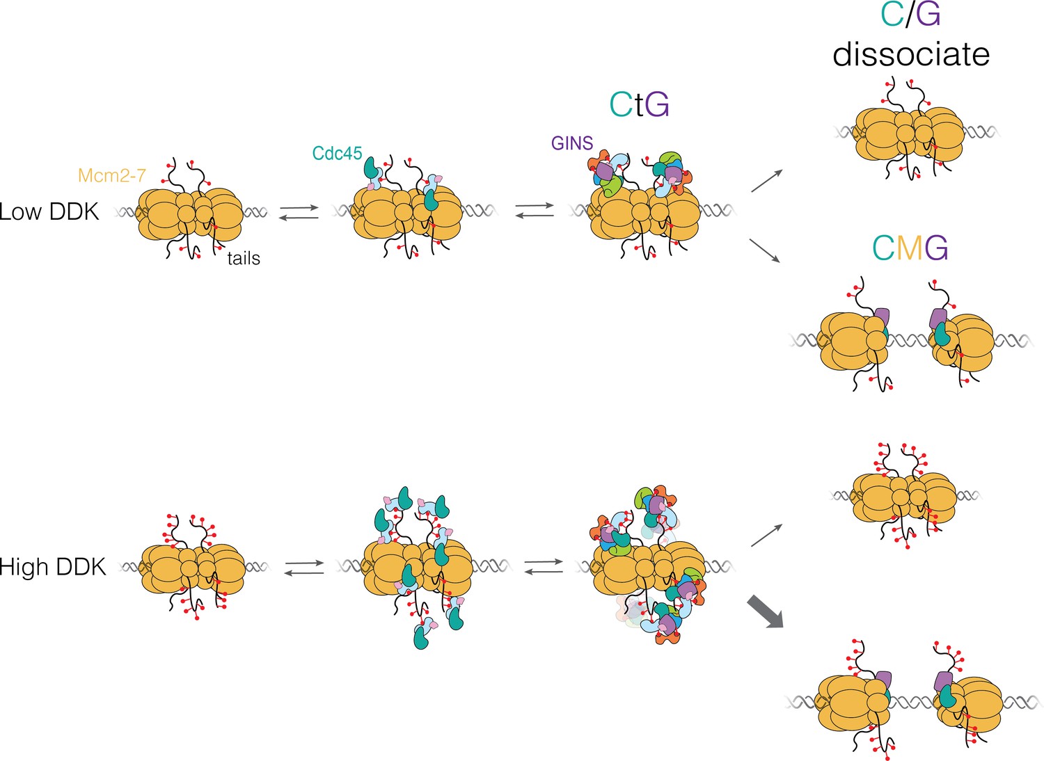
“Each time a eukaryotic cell divides (by mitosis) it must duplicate its chromosomal DNA exactly once to ensure that one full copy is passed to each resulting cell. Both under-replication or over-replication result in genome instability and disease or cell death. A key mechanism to prevent over-replication is the temporal separation of loading of the replicative DNA helicase [Mcm2-7] at origins of replication and activation of these same helicases during the cell division cycle.” Helicase loading is performed by the origin replication complex (ORC), a multi-subunit ATPase. In this study, Audra Amasino and Shalini Gupta from Steve Bell’s lab at MIT, working with Larry Friedman from the Gelles lab at Brandeis, define the mechanism by which cell cycle-dependent phosphorylation of the ORC inhibits helicase loading. Loading is a multi-step process and several steps are inhibited by phosphorylation, presumably helping to ensure that loading is completely suppressed during the S phase of the cell cycle during which the helicases are activated.

10.1073/pnas.2221484120

Amasino A., et al. Regulation of replication origin licensing by ORC phosphorylation reveals a two-step mechanism for Mcm2-7 ring closing. PNAS, 120, e2221484120 (2023).

RNA polymerase sliding on DNA can couple the transcription of nearby bacterial operons

Transcription initiation is arguably the single most important process for the regulation of gene expression in all organisms.  In bacteria, it is a widely accepted dogma that regulated transcription initiation at a promoter sequence controls only the adjacent transcription unit (i.e., a gene or operon).  In contrast, this paper demonstrates the feasibility of a new mechanism by which production of multiple RNAs from nearby operons is coupled to the binding of an individual RNA polymerase molecule to a promoter.

pnas.2301402120fig01

The new mechanism was hypothesized based on the recent discovery in vitro of a post-termination state of the bacterial RNA polymerase in which the polymerase slides randomly on DNA after terminating transcription of one RNA and can reinitiate transcription on a nearby promoter, producing another RNA. Despite these observations, it was unclear whether the hypothesized mechanism could operate efficiently over the time and distance scales necessary to couple nearby operons in a bacterial genome in vivo. Now-graduated Ph.D. student Debora Tenenbaum and collaborators tested this idea by developing a mathematical theory based on a diffusion-to-capture mechanism. The theory quantitatively predicts the efficacy of operon coupling in terms of rate constants that were previously unknown but which the authors measured in single-molecule biophysics experiments. This combination of theory and experiment shows that the mechanism operates on the length and time scales needed to function in bacterial genomes. The results suggest a generalized mechanism that couples the transcription of nearby operons and breaks the paradigm that each binding of RNAP to DNA can produce at most one messenger RNA.

10.1073/pnas.2301402120
Tenenbaum D., et al. RNA polymerase sliding on DNA can couple the transcription of nearby bacterial operons. PNAS, 120, e2301402120 (2023)

Recycling of bacterial RNA polymerase by the Swi2/Snf2 ATPase RapA

Molecular recycling is essential to gene expression.  In all organisms, after each RNA molecule is made, the RNA polymerase (RNAP) enzyme that made it must be reset and repositioned to make subsequent RNAs.  The steps involved in this RNAP recycling process are fully understood in any organism, not even in simple bacteria.

Ph.D. student Koe Inlow and collaborators studied the RapA enzyme, one of the only bacterial homologs of the large Swi2/Snf2 family of eukaryotic chromatin remodelers.  RapA has long been known as an abundant RNAP binding protein.  It has been proposed to function in RNAP recycling (and also in multiple other roles), but how it does so is unclear. Using multi-color single-molecule fluorescence microscopy, she made unprecedented direct observations in vitro of the dynamics of individual molecules of fluorescently labeled RNAP and RapA as they interacted with each other and with template DNA during and following transcript synthesis.  These studies show for the first time that RapA acts on a key intermediate in the transcription cycle: the recently discovered post-termination complex (PTC) in which RNAP slides along DNA and from which it can reinitiate transcription on nearby genes either sense or antisense relative to the prior round of transcript synthesis.  RapA thus competes against local reinitiation by RNAP sliding and is likely to promote replenishment of the global pool of free RNAP holoenzyme.  Further, the studies reveal that even tiny (nanomolar) concentrations of RapA efficiently use ATP hydrolysis to disassemble the PTC and uncover the essential features of the mechanism by which this removal occurs. These studies fill in the essential missing pieces in the current understanding of the events that occur after RNA is released and that enable RNAP recycling. Furthermore, we rationalize RapA genetics by explaining how RapA facilitates global transcriptional reprogramming as cells enter and leave stress conditions.

10.1073/pnas.2303849120
Inlow K., et al. Recycling of bacterial RNA polymerase by the Swi2/Snf2 ATPase RapA. PNAS 120, e2303849120 (2023)

Single-molecule analysis of actin filament debranching by cofilin and GMF

The Arp2/3 complex is a multi-component molecular machine that nucleates branched actin filament networks at the leading edge of cells to promote protrusion and at sites of endocytosis to drive membrane invagination. While the process of branched actin nucleation is now well understood (including at mechanistic and structural levels), what is less well understood is how the actin networks are subsequently debranched, or ‘pruned’. Debranching is an absolutely essential step in network remodeling and turnover, which is required for cell motility and endocytosis. The branched actin structures produced by Arp2/3 complex are kinetically stable, with spontaneous dissociation occurring only after tens of minutes to hours, whereas in vivo the branches dissociate in seconds. How is this achieved?

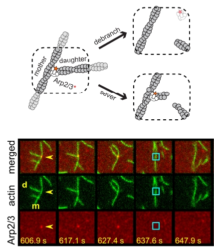
Two separate members of the larger ADF-homology family of proteins, glia maturation factor (GMF) and cofilin, have been implicated in promoting debranching. In this paper, Gelles lab member Johnson Chung, in collaboration with Jeff and with Bruce Goode from the Brandeis Biology Dept., used multi-wavelength single molecule florescence microscopy and quantitative kinetic analysis to define the mechanisms by which these proteins promote debranching.   Dr. Chung shows that “cofilin, like GMF, is an authentic debrancher independent of its filament-severing activity and that the debranching activities of the two proteins are additive. While GMF binds directly to the Arp2/3 complex, cofilin selectively accumulates on branch–junction daughter filaments in tropomyosin-decorated networks just prior to debranching events. Quantitative comparison of debranching rates with the known kinetics of cofilin–actin binding suggests that cofilin occupancy of a particular single actin site at the branch junction is sufficient to trigger debranching. In rare cases in which the order of departure could be resolved during GMF- or cofilin-induced debranching, the Arp2/3 complex left the branch junction bound to the pointed end of the daughter filament, suggesting that both GMF and cofilin can work by destabilizing the mother filament–Arp2/3 complex interface. Taken together, these observations suggest that GMF and cofilin promote debranching by distinct yet complementary mechanisms.”

10.1073/pnas.2115129119
Chung J, et al. Single-molecule analysis of actin filament debranching by cofilin and GMF.
PNAS,119, e2115129119 (2022)

Bayesian machine learning analysis of single-molecule fluorescence colocalization images

From Science at Brandeis: “Yerdos Ordabayev et al. in the Department of Biochemistry use Bayesian probabilistic programming to implement computer software “Tapqir” for analysis of colocalization single-molecule spectroscopy (CoSMoS) image data. CoSMoS is a tool widely used in vitro to study the biochemical and physical mechanisms of the protein and nucleic acid macromolecular “machines” that perform essential biological functions. In this method, formation and/or dissociation of molecular complexes is observed by single-molecule fluorescence microscopy as the colocalization of binder and target macromolecules each labeled with a different color of fluorescent dye. Despite the use of the method for over twenty years, reliable analysis of CoSMoS data remains a significant challenge to the effective and more widespread use of the technique.

This work describes a holistic causal probabilistic model of CoSMoS image data formation. This model is physics-based and includes realistic shot noise in fluorescent spots, camera noise, the size and shape of spots, and the presence of both specific and nonspecific binder molecules in the images. Most importantly, instead of yielding a binary spot-/no-spot determination, the algorithm calculates the probability of a colocalization event. Unlike alternative approaches, Tapqir does not require subjective threshold settings of parameters so they can be used effectively and accurately by non-expert analysts. The program is implemented in the state-of-the-art Python-based probabilistic programming language Pyro (open-sourced by Uber AI Labs in 2017), which enables efficient use of graphics processing unit (GPU)-based hardware for rapid parallel processing of data and facilitates future modifications to the model. Tapqir is free, open-source software. We envision that [the] program is likely to be adopted by researchers who use single-molecule colocalization methods to study a wide range of different biological systems.”

Yerdos is a postdoctoral fellow jointly advised by Profs. Douglas Theobald and Jeff Gelles.


10.7554/eLife.73860
Ordabayev Y.A., et al. Bayesian machine learning analysis of single-molecule fluorescence colocalization images
eLife, 11, e73860 (2022)

The endoplasmic reticulum chaperone BiP is a closure-accelerating cochaperone of Grp94

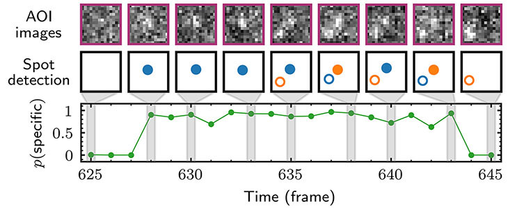
From the article: “The endoplasmic reticulum (ER) is the site at which secreted proteins (such as the hormone insulin) and membrane-bound proteins are folded. ATP-dependent chaperones within the ER help proteins fold. This study describes how two key ER chaperones, BiP and Grp94, work together at a molecular level. BiP binds to Grp94, which enables Grp94 to change conformation and hydrolyze ATP. In short, BiP provides a signal to switch on Grp94 conformational changes that are required to help other proteins fold. This finding helps explain how two chaperones can work together collaboratively in protein folding. Because BiP and Grp94 are members of highly conserved chaperone families, these findings may provide insight into chaperone-assisted protein folding beyond the ER.”  This project was a collaboration with members of Timothy Street‘s lab in the Brandeis Biochemistry Department.

10.1073/pnas.2118793119
Huang B., et al., The endoplasmic reticulum chaperone BiP is a closure-accelerating cochaperone of Grp94.
PNAS 119, e2118793119 (2022)

A helicase-tethered ORC flip enables bidirectional helicase loading

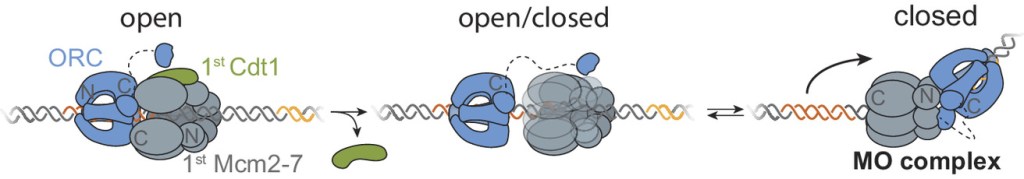
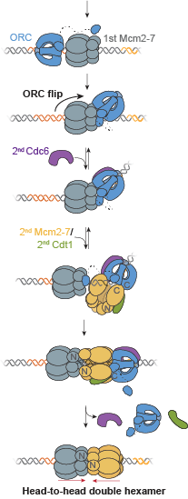
A key event in eukaryotic DNA replication is origin licensing in G1-phase, during which two Mcm2-7 replicative DNA helicases are loaded onto each origin DNA in an inactive, head-to-head fashion. Origin licensing marks every potential origin in a cell, and the opposing orientation of the loaded helicases ensures that they are poised to initiate bidirectional replication when the cell enters S-phase. Although it has long been known that the origin-recognition complex (ORC) binds origin DNA to direct helicase loading, the molecular mechanism by which two oppositely oriented helicases are loaded remains puzzling. Previous biochemical studies found evidence in support of a two-ORC mechanism for helicase loading wherein each of the two Mcm2-7 helicases are recruited by a separate, oppositely oriented ORC molecule. In contrast, single-molecule and cryo-EM approaches observed predominantly one ORC involved in helicase loading, but could not explain how a single ORC could load two oppositely oriented helicases.

In this paper, a collaboration with Steve Bell’s lab at MIT, Ph.D. student Shalini Gupta reconciles these seemingly contradictory observations. Using single-molecule fluorescence energy transfer (sm-FRET), she observed interactions in vitro between individual ORC molecules and the Mcm2-7 helicases in real time at two separate interfaces. In the large majority of instances, a single ORC molecule recruits both Mcm2-7 helicases through direct interactions. Between recruitment of the first and the second helicase, ORC ‘flips’ its orientation on DNA using a flexible protein tether to the first loaded Mcm2-7. This remarkable ORC inversion ensures that the two helicases are recruited via similar interactions, but in opposite orientations. The data define a complete, integrated pathway for helicase loading that resolves the apparent contradictions between previous observations. The tethered-flip mechanism provides a molecular explanation for how cells avoid the potentially damaging consequences of incompletely-formed helicase pairs at origins.

10.7554/eLife.74282
Gupta S., et al. A helicase-tethered ORC flip enables bidirectional helicase loading
eLife 10, e74282 (2021)

This article was the subject of an eLifeInsight article” by Bruce Stillman.

DDK regulates replication initiation by controlling the multiplicity of Cdc45-GINS binding to Mcm2-7

Schematic representation of DNA replication initiation Replication of chromosomal DNA in eukaryotes has two major stages.  Starting in the G1 phase of the cell cycle, double hexamers consisting of two copies of the Mcm2-7 replicative helicase are assembled at replication origins.   Later, in S phase, the two helicases are incorporated into two oppositely oriented CMG (Cdc45-Mcm2-7-GINS) complexes that each then form the core of a replisome.  Control of this “activation” step, which is triggered by the protein kinases DDK and S-CDK, is essential to ensure that each part of the genome is replicated once and only once in each cell cycle.

In this paper, Lorraine De Jesús-Kim together with collaborators from Steve Bell’s and Jeff Gelles’ labs used multi-wavelength single-molecule fluorescence colocation  (“CoSMoS”) methods to study in vitro the molecular mechanism of the activation process.  The journal’s acceptance summary notes that “The manuscript provides new and convincing evidence that a heretofore unknown intermediate state [called “CtG”] for replication start contains multiple copies of the GINS and Cdc45 proteins prior to initiation at each origin with one double hexamer of the MCM2-7 complex. The number of GINS and Cdc45 is determined by DDK phosphorylation of the MCM’s and the probability to create an active helicase (CMG) is increased with multiple numbers of the bound ancillary factors…. The single molecule studies and biochemistry are beautifully executed providing the evidence for such intermediates…. The addition of in vivo studies demonstrates that modulating the multiplicity of DDK phosphorylation (and proposed, CtG formation) has an impact on origin usage in cells.”Proposed model for Cdc45-Mcm2-7-GINS (CMG) formation

10.7554/eLife.65471
Kim L.D.J., et al., DDK regulates replication initiation by controlling the multiplicity of Cdc45-GINS binding to Mcm2-7.
eLife 10, e65471 (2021)

Dynamics of RNA polymerase II and elongation factor Spt4/5 recruitment during activator-dependent transcription

DNA transcription by RNA polymerase II (RNApII) is arguably the process most central to regulation of gene expression in eukaryotic organisms.  Regulated transcription requires the formation on DNA of molecular assemblies containing not only RNApII but also dozens of accessory proteins that play pivotal roles in the process.  While we know about the structures of some of these assemblies in atomic detail, quantitative understanding of the dynamics and pathways by which the assemblies interconvert and progress through this fundamental gene expression pathway is largely lacking.

In this study we report single-molecule fluorescence microscopy studies of transcription in yeast nuclear extract, for the first time visualizing and measuring the dynamics of activator-dependent recruitment of RNApII and the central elongation factor Spt4/5 to transcription complexes.  Grace Rosen (Jeff Gelles’ labortatory, Brandeis) , Inwha Baek (Steve, Buratowski’s lab, Harvard Medical School), and collaborators elucidated the kinetically significant steps in activated RNApII transcription initiation and show for the first time that Spt4/5 dynamics are tuned to the typical lifetimes of transcription elongation complexes.  In addition to these substantive results, our work represents an important methodological advance.  As the first application of the CoSMoS (co-localization single-molecule spectroscopy) technique to activated eukaryotic transcription, it demonstrates a general method for elucidating the correlated dynamic interactions of different components of the machinery with initiation and elongation transcription complexes.  The approach is likely to find further use in studies of the mechanistic features of RNApII transcription.

https://doi.org/10.1073/pnas.2011224117
Rosen, G.A., Baek, I., et al., Dynamics of RNA polymerase II and elongation factor Spt4/5 recruitment during activator-dependent transcription
PNAS 

Twinfilin bypasses assembly conditions and actin filament aging to drive barbed end depolymerization

The dynamic assembly, remodeling, and turnover of actin networks drives cellular processes
ranging from cell motility, endocytosis, and phagocytosis to cell division, cell and tissue
morphogenesis, and neuronal pathfinding. Here, we describe a new actin regulatory activity that changes understanding of how actin networks can be turned over. In a collaborative project with Bruce Goode’s lab, postdocs Shashank Shekhar and Greg Hoeprich used microfluidics-assisted total internal reflection fluorescence (TIRF) microscopy to show that mammalian twinfilin, an evolutionary conserved ADF/cofilin-homology protein, accelerates depolymerization at newly-assembled (ADP-Pi) but not older (ADP) actin filaments, even under assembly-promoting conditions (i.e., at G-actin concentrations above the critical concentration). Our data suggest that twinfilin molecules interact processively with the barbed end of the filament as it shrinks, blocking ATP-actin subunit addition while allowing ADP-Pi subunit dissociation. These novel activities of twinfilin reveal that cells have machinery that can bypass the normal filament aging process and induce the depolymerization of barbed ends as needed. These results may explain known genetic interactions between twinfilin and cofilin, and localization of twinfilin to the tips of filopodia and stereocilia, where actin filament barbed ends are clustered.

[ensemblevideo contentid=94fc0a2d-eaad-4ccb-84ac-7ebf799ba3ec autoplay=true]

10.1083/jcb.202006022
Shekhar S, et al., Twinfilin bypasses assembly conditions and actin filament aging to drive barbed end depolymerization.
Journal of Cell Biology
220, e202006022 (2021)