
We are delighted to have Biochemistry and Biophysics student Ziwei Wang as the newest member of the Gelles Lab. Welcome, Ziwei!
Grant renewal: Single-molecule visualization of transcription regulation mechanisms
The National Institute of General Medical Sciences, National Institutes of![]() Health, recently awarded a four-year renewal of our grant “Single-molecule visualization of transcription regulation mechanisms”. This award funds Gelles Lab research on the molecular bases of DNA transcription and gene regulation in both bacteria and in eukaryotic organisms.
Health, recently awarded a four-year renewal of our grant “Single-molecule visualization of transcription regulation mechanisms”. This award funds Gelles Lab research on the molecular bases of DNA transcription and gene regulation in both bacteria and in eukaryotic organisms.
“Single-molecule visualization of a formin-capping protein ‘decision complex’ at the actin filament barbed end”
[ensemblevideo contentid=Z33TDbsofEW7ofOXsSti8w autoplay=true]Regulation of actin filament length is a central process by which eukaryotic cells control the shape, architecture, and dynamics of their actin networks. This regulation plays a fundamental role in cell motility, morphogenesis, and a host of processes specific to particular cell types. This paper by recently graduated Ph.D. student Jeffrey Bombardier and collaborators resolves the long-standing mystery of how formins and capping protein work in concert and antagonistically to control actin filament length. Bombardier used the CoSMoS multi-wavelength single-molecule fluorescence microscopy technique to to discover and characterize a novel tripartite complex formed by a formin, capping protein, and the actin filament barbed end. Quantitative analysis of the kinetic mechanism showed that this complex is the essential intermediate and decision point in converting a growing formin-bound filament into a static capping protein-bound filament, and the reverse. Interestingly, the authors show that “mDia1 displaced from the barbed end by CP can randomly slide along the filament and later return to the barbed end to re-form the complex.” The results define the essential features of the molecular mechanism of filament length regulation by formin and capping protein; this mechanism predicts several new ways by which cells are likely to couple upstream regulatory inputs to filament length control.
10.1038/NCOMMS9707Single-molecule visualization of a formin-capping protein ‘decision complex’ at the actin filament barbed end
Jeffrey P. Bombardier, Julian A. Eskin, Richa Jaiswal, Ivan R. Corrêa, Jr., Ming-Qun Xu, Bruce L. Goode, and Jeff Gelles
Nature Communications 6:8707 (2015)
Resources: The capping protein expression plasmid described in this article is available from Addgene.
Readers interested in this subject should also see a related article by Shekhar et al published simultaneously in the same journal. We are grateful to the authors of that article for coordinating submission so that the two articles were published together.
BioTechniques highlights single-molecule enzymology
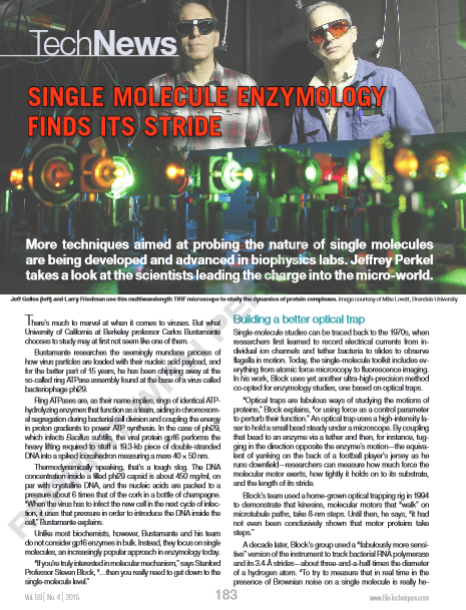 BioTechniques published a TechNews feature by Jeffrey Perkel that discusses advances in single-molecule studies of enzymes from a number of labs, including ours. There is a nice description of Simina Ticau’s paper on licensing of eukaryotic DNA replication origins.
Perkel, J.
Single Molecule Enzymology Finds its Stride
BioTechniques 59:183–187 (2015).
“Multi-wavelength single-molecule fluorescence analysis of transcription mechanisms”
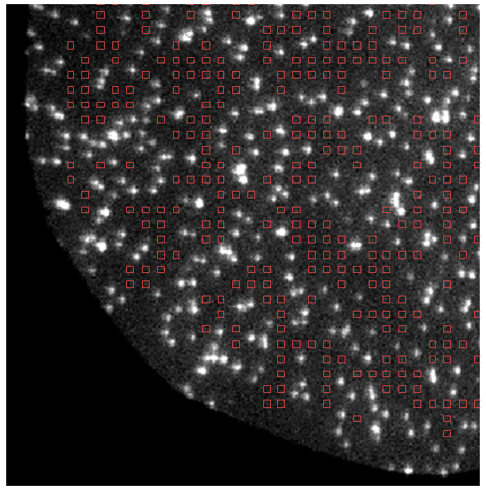 In single-molecule colocalization (“CoSMoS”) experiments, “the most challenging aspects of the technique are often not the preparation of the molecules or making the microscope observations, but in analyzing the resulting data. Data analysis has two primary challenges. First, one is studying inherently stochastic processes (thermally driven reactions of single molecules), so that analysis is inherently statistical. Second, the number of photons that can be emitted by a single fluorophore is limited by photobleaching, so images often have low signal-to-noise ratios, making discrimination of real signals from noise a challenge.” In this article, Larry Friedman provides a step-by-step description the the procedures he has developed to reliably and efficiently analyze CoSMoS data. While the examples discussed in the paper focus on analysis of transcription mechanisms, the methods should be applicable to CoSMoS studies on a broad range of biological systems.
Multi-wavelength single-molecule fluorescence analysis of transcription mechanisms.
Larry J. Friedman and Jeff Gelles
Methods 86, 27–36 (2015)
Resources: Computer software that implements methods described in this article is available here.
“Single-molecule imaging of a three-component ordered actin disassembly mechanism”
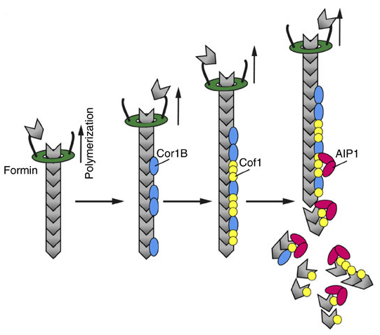 “The mechanisms by which cells destabilize and rapidly disassemble filamentous actin networks have remained elusive; however, Coronin, Cofilin and AIP1 have been implicated in this process.” Silvia Jansen from Bruce Goode’s lab and co-authors showed in this study “using multi-wavelength single-molecule fluorescence imaging…that mammalian Cor1B, Cof1 and AIP1 work in concert through a temporally ordered pathway to induce highly efficient severing and disassembly of actin filaments. Cor1B binds to filaments first, and dramatically accelerates the subsequent binding of Cof1, leading to heavily decorated, stabilized filaments. Cof1 in turn recruits AIP1, which rapidly triggers severing and remains bound to the newly generated barbed ends. New growth at barbed ends generated by severing was blocked specifically in the presence of all three proteins. This activity enabled us to reconstitute and directly visualize single actin filaments being rapidly polymerized by formins at their barbed ends while simultanteously being stochastically severed and capped along their lengths, and disassembled from their pointed ends.”
Single-molecule imaging of a three-component ordered actin disassembly mechanism
Silvia Jansen, Agnieszka Collins, Samantha M. Chin, Casey A. Ydenberg, Jeff Gelles. Bruce L. Goode
Nature Communications 6, 7202 (2015)
“Single-Molecule Studies of Origin Licensing Reveal Mechanisms Ensuring Bidirectional Helicase Loading”
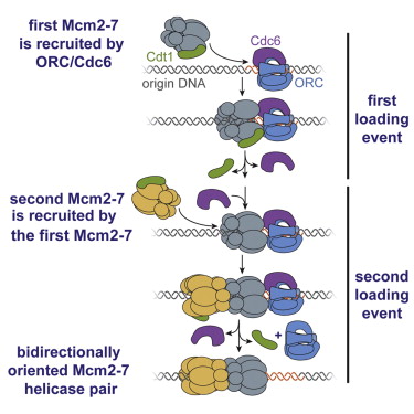 In this study, a collaboration between the Gelles lab and Stephen P. Bell’s lab at MIT, Simina Ticau and co-authors studied the mechanism of “licensing”, the initial process in setting up DNA replication in eukaryotic cells. “Licensing is loading of the ring-shaped Mcm2-7 helicase around DNA origins of replication. During loading, Cdc6, Cdt1, and the origin-recognition complex (ORC) assemble two heterohexameric Mcm2-7 complexes into a head-to-head double hexamer that facilitates bidirectional replication initiation. Using multi-wavelength single-molecule fluorescence to monitor the events of helicase loading, we demonstrate that double-hexamer formation is the result of sequential loading of individual Mcm2-7 complexes. Loading of each Mcm2-7 molecule involves the ordered association and dissociation of distinct Cdc6 and Cdt1 proteins. In contrast, one ORC molecule directs loading of both helicases in each double hexamer. Based on single-molecule FRET, arrival of the second Mcm2-7 results in rapid double-hexamer formation that anticipates Cdc6 and Cdt1 release, suggesting that Mcm-Mcm interactions recruit the second helicase. Our findings reveal the complex protein dynamics that coordinate helicase loading and indicate that distinct mechanisms load the oppositely oriented helicases that are central to bidirectional replication initiation.”
Ticau, S., Friedman, L.J., Ivica, N.A., Gelles, J. & Bell, S.P.
Single-Molecule Studies of Origin Licensing Reveal Mechanisms Ensuring Bidirectional Helicase Loading.
Cell 161, 513-525 (2015).
This article was featured in a Cell Preview “Single-Molecule Visualization of MCM2-7 DNA Loading: Seeing Is Believing” by Gheorghe Chisto and Johannes C. Walter. Cell 161, 429-430 (2015) 10.1016/j.cell.2015.04.006. It was also discussed in
“Single Molecule Enzymology Finds its Stride” by Jeffrey Perkel. BioTechniques 59:183–187 (2015) 10.2144/000114337.
New lab members and summer students
Welcome to new students Matt Chamberlain, Grace Rosen, and Yanding Zhao!
“Single-molecule studies of actin assembly and disassembly factors”
 This new Methods in Enzymology chapter by Ben Smith, Jeff Gelles, and Bruce Goode “describe[s] techniques for acquisition and analysis of single-molecule data, applied to the novel challenges of studying the filament assembly and disassembly activities of actin-associated proteins in vitro. We discuss the advantages of single-molecule analysis in directly visualizing the order of molecular events, measuring the kinetic rates of filament binding and dissociation, and studying the coordination among multiple factors. The methods described… complement traditional biochemical approaches in elucidating actin-regulatory mechanisms in reconstituted filamentous networks.”
Methods Enzymol. 2014;540:95-117.
Single-molecule studies of actin assembly and disassembly factors.
Smith BA, Gelles J, Goode BL
