Category Archives: Publications

Dynamics of GreB-RNA polymerase interaction

In living cells, messenger RNAs are not manufactured by RNA polymerases (RNAPs) functioning alone.  Instead, RNA synthesis is carried out collectively by RNAP together with accessory proteins that associate with the RNAP-containing transcripti
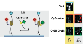
on elongation complex and modulate its activity.  In this paper, Larry Tetone, Larry Friedman, and Melissa Osborne, along with their collaborators from the Gelles and Landick labs, used multi-wavelength single-molecule fluorescence methods to for the first time directly observe the dynamic binding and dissociation of an accessory protein with an RNAP during active transcript elongation.  The protein, GreB, is important for transcript proofreading in E. coli and other bacteria and is a functional analog of the TFIIS protein in eaukaryotes.  “Unexpectedly,” the authors report, “GreB was not selectively recruited to RNAPs requiring its transcript proofreading function. Instead, GreB transiently bound to multiple types of complexes, eventually finding via random search RNAPs that require its activity. The observations suggest a paradigm by which a regulator can act while minimizing obstruction of a binding site that must be shared with other proteins.”

10.1073/pnas.1616525114
Dynamics of GreB-RNA polymerase interaction allow a proofreading accessory protein to patrol for transcription complexes needing rescue
Larry E. Tetone, Larry J. Friedman, Melisa L. Osborne, Harini Ravi, Scotty Kyzer, Sarah K. Stumper, Rachel A. Mooney, Robert Landick, and Jeff Gelles
PNAS (2017) 114:E1081-E1090.

Resources:
New plasmids reported in this article can be obtained from Addgene

“The dimerization equilibrium of a ClC Cl−/H+ antiporter in lipid bilayers”

The physical forces that drive oligomerization of soluble proteins are well understood and have been extensively studied.  For proteins with transmembrane domains — transport enzymes, for example — oligomerization is often essential for function but its physical basis is less clear.  In this project,  Janice Robertson devised a new method based on liposome extrusion and single-molecule fluorescence photobleaching analysis to accurately measure the dimer association free energy of a ClC-type chloride ion/hydrogen ion antiporter.  (Janice started this work when she was a postdoc in Chris Miller’s lab at Brandeis and later completed the project in her own  lab at the University of Iowa.)  The study reveals that ClC-ec1 “is one of the strongest membrane protein complexes measured so far, and introduces it as new type of dimerization model to investigate the physical forces that drive membrane protein association in membranes.”

10.7554/eLife.17438
The dimerization equilibrium of a ClC Cl−/H+ antiporter in lipid bilayers
Rahul Chadda, Venkatramanan Krishnamani, Kacey Mersch, Jason Wong, Marley Brimberry, Ankita Chadda, Ludmila Kolmakova-Partensky, Larry J Friedman, Jeff Gelles, and Janice L Robertson
eLife (2016) 5:e17438

“Single molecule analysis reveals reversible and irreversible steps during spliceosome activation”

“The spliceosome is a complex molecular machine, composed of small nuclear ribonucleoproteins (snRNPs) and accessory proteins, that excises introns from precursor messenger RNAs (pre-mRNAs).  After assembly, the spliceosome is activated for catalysis by rearrangement of subunits to form an active site.” This study used multi-wavelength single-molecule fluorescence (“CoSMoS”) techniques to elucidate the mechanism of budding yeast spliceosome activation.  Activation turns out to be unexpectedly dynamic and variable: some spliceosomes  take multiple attempts to activate and the pathway contains both reversible and irreversible steps.  Strikingly, ATP powers both steps that drive the process forward toward splicing and well as reverse steps that diassemble intermediates to allow subsequent re-attempts at activation. These findings give new insight into how the efficiency and fidelity of pre-mRNA splicing is maintained.Predominant spliceosome activation pathway

The scientific project in this paper was initiated by Aaron Hoskins during his postdoctoral work in Melissa Moore’s and Jeff Gelles’ labs, but it was brought to fruition by Aaron and Margaret Rodgers working in Aaron’s lab at Univ. Wisconsin, Madison.

10.7554/eLife.14166
Single molecule analysis reveals reversible and irreversible steps during spliceosome activation
Aaron A. Hoskins Margaret L. Rodgers , Larry J. Friedman , Jeff Gelles , Melissa J. Moore
eLife (2016) 5:e14166

“Bacterial RNA polymerase can retain σ70 throughout transcription”

“In all kingdoms of life, gene transcription is not carried out by RNA polymerase enzymes alone.” Instead, accessory proteins ride along with RNA polymerase molecules as the latter move along a gene, regulating their biological function and controlling gene expression. However, in no cases is the kinetic mechanism of such elongation regulation quantitatively understood.

Sigma proteins are known to be regulators of bacterial transcription initiation. However, previous work suggested that σ70 is present on some transcription elongation complexes, although the extent to which it is retained from initiation, how long it remains attached, and its consequences for transcription regulation were unclear. In this study, Tim Harden and his collaborators used a novel multi-wavelength single-molecule fluorescence microscopy approach to directly observe and quantitatively characterize the dynamic interactions of the σ70 protein with bacterial RNA polymerase molecules in vitro during active RNA synthesis. Harden is a Brandeis Physics Ph.D. student who is jointly advised by Jeff Gelles and Jane Kondev.  The study demonstrates by direct observation that actively elongating polymerase molecules can retain σ70 from initiation into the elongation phase of transcription; shows that retained σ70 subunits dissociate so slowly that most are still present on the elongation complex at the end of a long gene; and proves that only the subpopulation of elongating polymerases with bound σ70 recognize a class of transcriptional pause sequences which in some contexts play a well-established role in regulating gene expression.hardenfig

More generally, this study provides the first quantitative framework that defines the post-initiation roles of σ70, information that is essential to the understanding of global transcription regulation in bacteria. Furthermore, the work demonstrates a general method for elucidating the dynamic interactions of transcription factors with active elongation complexes; this method has broad application in both prokaryotic and eukaryotic transcription biology.

10.1073/pnas.1513899113
Bacterial RNA polymerase can retain σ70 throughout transcription
Timothy T. Harden, Christopher D. Wells, Larry J. Friedman, Robert Landick, Ann Hochschild, Jane Kondev, and Jeff Gelles
PNAS (2016) 113:602-607

Resources: Plasmids described in this article are available from Addgene.

“Single-molecule visualization of a formin-capping protein ‘decision complex’ at the actin filament barbed end”

[ensemblevideo contentid=Z33TDbsofEW7ofOXsSti8w autoplay=true]Regulation of actin filament length is a central process by which eukaryotic cells control the shape, architecture, and dynamics of their actin networks. This regulation plays a fundamental role in cell motility, morphogenesis, and a host of processes specific to particular cell types. This paper by recently graduated Ph.D. student Jeffrey Bombardier and collaborators resolves the long-standing mystery of how formins and capping protein work in concert and antagonistically to control actin filament length. Bombardier used the CoSMoS multi-wavelength single-molecule fluorescence microscopy technique to to discover and characterize a novel tripartite complex formed by a formin, capping protein, and the actin filament barbed end. Quantitative analysis of the kinetic mechanism showed that this complex is the essential intermediate and decision point in converting a growing formin-bound filament into a static capping protein-bound filament, and the reverse. Interestingly, the authors show that “mDia1 displaced from the barbed end by CP can randomly slide along the filament and later return to the barbed end to re-form the complex.” The results define the essential features of the molecular mechanism of filament length regulation by formin and capping protein; this mechanism predicts several new ways by which cells are likely to couple upstream regulatory inputs to filament length control.

10.1038/NCOMMS9707
Single-molecule visualization of a formin-capping protein ‘decision complex’ at the actin filament barbed end
Jeffrey P. Bombardier, Julian A. Eskin, Richa Jaiswal, Ivan R. Corrêa, Jr., Ming-Qun Xu, Bruce L. Goode, and Jeff Gelles
Nature Communications  6:8707 (2015)

Resources: The capping protein expression plasmid described in this article is available from Addgene.

Readers interested in this subject should also see a related article by Shekhar et al published simultaneously in the same journal.  We are grateful to the authors of that article for coordinating submission so that the two articles were published together.

“Multi-wavelength single-molecule fluorescence analysis of transcription mechanisms”

Figure9In single-molecule colocalization (“CoSMoS”) experiments, “the most challenging aspects of the technique are often not the preparation of the molecules or making the microscope observations, but in analyzing the resulting data. Data analysis has two primary challenges. First, one is studying inherently stochastic processes (thermally driven reactions of single molecules), so that analysis is inherently statistical. Second, the number of photons that can be emitted by a single fluorophore is limited by photobleaching, so images often have low signal-to-noise ratios, making discrimination of real signals from noise a challenge.”  In this article, Larry Friedman provides a step-by-step description the the procedures he has developed to reliably and efficiently analyze CoSMoS data.  While the examples discussed in the paper focus on analysis of transcription mechanisms, the methods should be applicable to CoSMoS studies on a broad range of biological systems.

10.1016/j.ymeth.2015.05.026
Multi-wavelength single-molecule fluorescence analysis of transcription mechanisms.
Larry J. Friedman and Jeff Gelles
Methods 86, 27–36 (2015)

Resources: Computer software that implements methods described in this article is available here.

“Single-molecule imaging of a three-component ordered actin disassembly mechanism”

sj2015“The mechanisms by which cells destabilize and rapidly disassemble filamentous actin networks have remained elusive; however, Coronin, Cofilin and AIP1 have been implicated in this process.”   Silvia Jansen from Bruce Goode’s lab and co-authors showed in this study “using multi-wavelength single-molecule fluorescence imaging…that mammalian Cor1B, Cof1 and AIP1 work in concert through a temporally ordered pathway to induce highly efficient severing and disassembly of actin filaments. Cor1B binds to filaments first, and dramatically accelerates the subsequent binding of Cof1, leading to heavily decorated, stabilized filaments. Cof1 in turn recruits AIP1, which rapidly triggers severing and remains bound to the newly generated barbed ends. New growth at barbed ends generated by severing was blocked specifically in the presence of all three proteins. This activity enabled us to reconstitute and directly visualize single actin filaments being rapidly polymerized by formins at their barbed ends while simultanteously being stochastically severed and capped along their lengths, and disassembled from their pointed ends.”

10.1038/ncomms8202
Single-molecule imaging of a three-component ordered actin disassembly mechanism
Silvia Jansen, Agnieszka Collins, Samantha M. Chin, Casey A. Ydenberg, Jeff Gelles. Bruce L. Goode
Nature Communications 6, 7202 (2015)

“Single-Molecule Studies of Origin Licensing Reveal Mechanisms Ensuring Bidirectional Helicase Loading”

Fig 7finalIn this study, a collaboration between the Gelles lab and Stephen P. Bell’s lab at MIT, Simina Ticau and co-authors studied the mechanism of “licensing”, the initial process in setting up DNA replication in eukaryotic cells. “Licensing is loading of the ring-shaped Mcm2-7 helicase around DNA origins of replication. During loading, Cdc6, Cdt1, and the origin-recognition complex (ORC) assemble two heterohexameric Mcm2-7 complexes into a head-to-head double hexamer that facilitates bidirectional replication initiation. Using multi-wavelength single-molecule fluorescence to monitor the events of helicase loading, we demonstrate that double-hexamer formation is the result of sequential loading of individual Mcm2-7 complexes. Loading of each Mcm2-7 molecule involves the ordered association and dissociation of distinct Cdc6 and Cdt1 proteins. In contrast, one ORC molecule directs loading of both helicases in each double hexamer. Based on single-molecule FRET, arrival of the second Mcm2-7 results in rapid double-hexamer formation that anticipates Cdc6 and Cdt1 release, suggesting that Mcm-Mcm interactions recruit the second helicase. Our findings reveal the complex protein dynamics that coordinate helicase loading and indicate that distinct mechanisms load the oppositely oriented helicases that are central to bidirectional replication initiation.”

10.1016/j.cell.2015.03.012
Ticau, S., Friedman, L.J., Ivica, N.A., Gelles, J. & Bell, S.P.
Single-Molecule Studies of Origin Licensing Reveal Mechanisms Ensuring Bidirectional Helicase Loading.
Cell 161, 513-525 (2015).

This article was featured in a Cell Preview “Single-Molecule Visualization of MCM2-7 DNA Loading: Seeing Is Believing” by Gheorghe Chisto and Johannes C. Walter. Cell 161, 429-430 (2015) 10.1016/j.cell.2015.04.006.  It was also discussed in
Single Molecule Enzymology Finds its Stride” by Jeffrey Perkel. BioTechniques 59:183–187 (2015) 10.2144/000114337.

“Single-molecule studies of actin assembly and disassembly factors”

micromirror-tirfThis new Methods in Enzymology chapter by Ben Smith, Jeff Gelles, and Bruce Goode “describe[s] techniques for acquisition and analysis of single-molecule data, applied to the novel challenges of studying the filament assembly and disassembly activities of actin-associated proteins in vitro. We discuss the advantages of single-molecule analysis in directly visualizing the order of molecular events, measuring the kinetic rates of filament binding and dissociation, and studying the coordination among multiple factors. The methods described… complement traditional biochemical approaches in elucidating actin-regulatory mechanisms in reconstituted filamentous networks.”

doi: 10.1016/B978-0-12-397924-7.00006-6 Pubmed Central
Methods Enzymol. 2014;540:95-117.
Single-molecule studies of actin assembly and disassembly factors.
Smith BA, Gelles J, Goode BL