In January, Jeff gave a lecture on Molecular Computers at the Center of Living Cells at the Wheeler Opera House in Aspen, Colorado. This lecture, sponsored by the Aspen Center for Physics and part of the 2017 Maggie & Nick DeWolf Physics Lecture Series, was intended to introduce members of the general public to recent developments in single-molecule biophysics.
Author Archives: Brandeis ITS
“The dimerization equilibrium of a ClC Cl−/H+ antiporter in lipid bilayers”
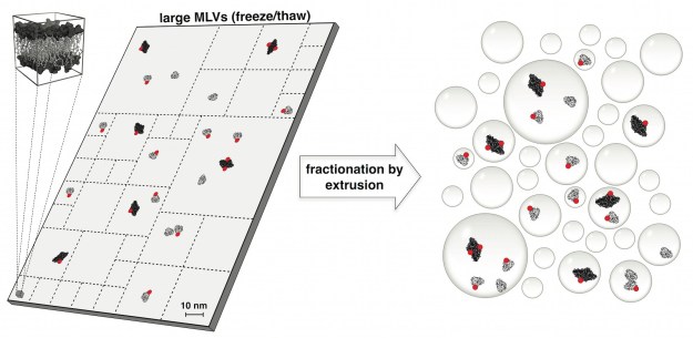
The physical forces that drive oligomerization of soluble proteins are well understood and have been extensively studied. For proteins with transmembrane domains — transport enzymes, for example — oligomerization is often essential for function but its physical basis is less clear. In this project, Janice Robertson devised a new method based on liposome extrusion and single-molecule fluorescence photobleaching analysis to accurately measure the dimer association free energy of a ClC-type chloride ion/hydrogen ion antiporter. (Janice started this work when she was a postdoc in Chris Miller’s lab at Brandeis and later completed the project in her own lab at the University of Iowa.) The study reveals that ClC-ec1 “is one of the strongest membrane protein complexes measured so far, and introduces it as new type of dimerization model to investigate the physical forces that drive membrane protein association in membranes.”

The dimerization equilibrium of a ClC Cl−/H+ antiporter in lipid bilayers
Rahul Chadda, Venkatramanan Krishnamani, Kacey Mersch, Jason Wong, Marley Brimberry, Ankita Chadda, Ludmila Kolmakova-Partensky, Larry J Friedman, Jeff Gelles, and Janice L Robertson
eLife (2016) 5:e17438
Timothy Harden, Ph.D.
Congratulations! to Tim Harden, who successfully defended his Ph.D. dissertation in Physics with an additional specialization in Quantitative Biology. Tim was jointly advised by Jeff Gelles and Jane Kondev. He is now a Postdoctoral Fellow in Angela DePace’s lab at Harvard Medical School.

L-to-R: Jeff, Tim, Jane
“Single molecule analysis reveals reversible and irreversible steps during spliceosome activation”
“The spliceosome is a complex molecular machine, composed of small nuclear ribonucleoproteins (snRNPs) and accessory proteins, that excises introns from precursor messenger RNAs (pre-mRNAs). After assembly, the spliceosome is activated for catalysis by rearrangement of subunits to form an active site.” This study used multi-wavelength single-molecule fluorescence (“CoSMoS”) techniques to elucidate the mechanism of budding yeast spliceosome activation. Activation turns out to be unexpectedly dynamic and variable: some spliceosomes take multiple attempts to activate and the pathway contains both reversible and irreversible steps. Strikingly, ATP powers both steps that drive the process forward toward splicing and well as reverse steps that diassemble intermediates to allow subsequent re-attempts at activation. These findings give new insight into how the efficiency and fidelity of pre-mRNA splicing is maintained.
The scientific project in this paper was initiated by Aaron Hoskins during his postdoctoral work in Melissa Moore’s and Jeff Gelles’ labs, but it was brought to fruition by Aaron and Margaret Rodgers working in Aaron’s lab at Univ. Wisconsin, Madison.
10.7554/eLife.14166Single molecule analysis reveals reversible and irreversible steps during spliceosome activation
Aaron A. Hoskins Margaret L. Rodgers , Larry J. Friedman , Jeff Gelles , Melissa J. Moore
eLife (2016) 5:e14166
Radcliffe Fellow
During Jeff’s sabbatical in 2016-17, he will be a Fellow at the Radcliffe Institute for Advanced Study at Harvard University, working on a project to study eukaryotic mRNA synthesis and matuaration mechanisms using single-molecule fluorescence methods. In addition to this new project he will also be spending time each week at Brandeis working with students and other scientists on the lab’s ongoing projects including those supported by NIH and the Mathers Foundation. (The lab will be accepting Ph.D. students for rotation projects during 2016-17.)
Grant renewal: Molecular mechanisms coordinating the actin and microtubule cytoskeletons
The National Institute of General Medical Sciences, National Institutes of Health, has awarded a four-year renewal of “Molecular mechanisms coordinating the actin and microtubule cytoskeletons”. This grant funds a joint research project of Jeff Gelles’ and Bruce Goode’s labs at Brandeis. The project is to determine the molecular processes by which microtubule and actin networks interact to control the architecture and dynamics of eukaryotic cells. The project has resulted in numerous publications co-authored by members of the two labs, and we are grateful for the opportunity to continue this research.
“Bacterial RNA polymerase can retain σ70 throughout transcription”
“In all kingdoms of life, gene transcription is not carried out by RNA polymerase enzymes alone.” Instead, accessory proteins ride along with RNA polymerase molecules as the latter move along a gene, regulating their biological function and controlling gene expression. However, in no cases is the kinetic mechanism of such elongation regulation quantitatively understood.
Sigma proteins are known to be regulators of bacterial transcription initiation. However, previous work suggested that σ70 is present on some transcription elongation complexes, although the extent to which it is retained from initiation, how long it remains attached, and its consequences for transcription regulation were unclear. In this study, Tim Harden and his collaborators used a novel multi-wavelength single-molecule fluorescence microscopy approach to directly observe and quantitatively characterize the dynamic interactions of the σ70 protein with bacterial RNA polymerase molecules in vitro during active RNA synthesis. Harden is a Brandeis Physics Ph.D. student who is jointly advised by Jeff Gelles and Jane Kondev. The study demonstrates by direct observation that actively elongating polymerase molecules can retain σ70 from initiation into the elongation phase of transcription; shows that retained σ70 subunits dissociate so slowly that most are still present on the elongation complex at the end of a long gene; and proves that only the subpopulation of elongating polymerases with bound σ70 recognize a class of transcriptional pause sequences which in some contexts play a well-established role in regulating gene expression.
More generally, this study provides the first quantitative framework that defines the post-initiation roles of σ70, information that is essential to the understanding of global transcription regulation in bacteria. Furthermore, the work demonstrates a general method for elucidating the dynamic interactions of transcription factors with active elongation complexes; this method has broad application in both prokaryotic and eukaryotic transcription biology.
10.1073/pnas.1513899113Bacterial RNA polymerase can retain σ70 throughout transcription
Timothy T. Harden, Christopher D. Wells, Larry J. Friedman, Robert Landick, Ann Hochschild, Jane Kondev, and Jeff Gelles
PNAS (2016) 113:602-607
Resources: Plasmids described in this article are available from Addgene.
New lab member: Ziwei Wang
We are delighted to have Biochemistry and Biophysics student Ziwei Wang as the newest member of the Gelles Lab. Welcome, Ziwei!
Grant renewal: Single-molecule visualization of transcription regulation mechanisms
The National Institute of General Medical Sciences, National Institutes of
Health, recently awarded a four-year renewal of our grant “Single-molecule visualization of transcription regulation mechanisms”. This award funds Gelles Lab research on the molecular bases of DNA transcription and gene regulation in both bacteria and in eukaryotic organisms.
“Single-molecule visualization of a formin-capping protein ‘decision complex’ at the actin filament barbed end”
[ensemblevideo contentid=Z33TDbsofEW7ofOXsSti8w autoplay=true]Regulation of actin filament length is a central process by which eukaryotic cells control the shape, architecture, and dynamics of their actin networks. This regulation plays a fundamental role in cell motility, morphogenesis, and a host of processes specific to particular cell types. This paper by recently graduated Ph.D. student Jeffrey Bombardier and collaborators resolves the long-standing mystery of how formins and capping protein work in concert and antagonistically to control actin filament length. Bombardier used the CoSMoS multi-wavelength single-molecule fluorescence microscopy technique to to discover and characterize a novel tripartite complex formed by a formin, capping protein, and the actin filament barbed end. Quantitative analysis of the kinetic mechanism showed that this complex is the essential intermediate and decision point in converting a growing formin-bound filament into a static capping protein-bound filament, and the reverse. Interestingly, the authors show that “mDia1 displaced from the barbed end by CP can randomly slide along the filament and later return to the barbed end to re-form the complex.” The results define the essential features of the molecular mechanism of filament length regulation by formin and capping protein; this mechanism predicts several new ways by which cells are likely to couple upstream regulatory inputs to filament length control.
10.1038/NCOMMS9707Single-molecule visualization of a formin-capping protein ‘decision complex’ at the actin filament barbed end
Jeffrey P. Bombardier, Julian A. Eskin, Richa Jaiswal, Ivan R. Corrêa, Jr., Ming-Qun Xu, Bruce L. Goode, and Jeff Gelles
Nature Communications 6:8707 (2015)
Resources: The capping protein expression plasmid described in this article is available from Addgene.
Readers interested in this subject should also see a related article by Shekhar et al published simultaneously in the same journal. We are grateful to the authors of that article for coordinating submission so that the two articles were published together.
