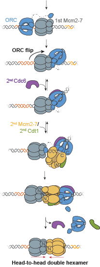
 A key event in eukaryotic DNA replication is origin licensing in G1-phase, during which two Mcm2-7 replicative DNA helicases are loaded onto each origin DNA in an inactive, head-to-head fashion. Origin licensing marks every potential origin in a cell, and the opposing orientation of the loaded helicases ensures that they are poised to initiate bidirectional replication when the cell enters S-phase. Although it has long been known that the origin-recognition complex (ORC) binds origin DNA to direct helicase loading, the molecular mechanism by which two oppositely oriented helicases are loaded remains puzzling. Previous biochemical studies found evidence in support of a two-ORC mechanism for helicase loading wherein each of the two Mcm2-7 helicases are recruited by a separate, oppositely oriented ORC molecule. In contrast, single-molecule and cryo-EM approaches observed predominantly one ORC involved in helicase loading, but could not explain how a single ORC could load two oppositely oriented helicases.
In this paper, a collaboration with Steve Bell’s lab at MIT, Ph.D. student Shalini Gupta reconciles these seemingly contradictory observations. Using single-molecule fluorescence energy transfer (sm-FRET), she observed interactions in vitro between individual ORC molecules and the Mcm2-7 helicases in real time at two separate interfaces. In the large majority of instances, a single ORC molecule recruits both Mcm2-7 helicases through direct interactions. Between recruitment of the first and the second helicase, ORC ‘flips’ its orientation on DNA using a flexible protein tether to the first loaded Mcm2-7. This remarkable ORC inversion ensures that the two helicases are recruited via similar interactions, but in opposite orientations. The data define a complete, integrated pathway for helicase loading that resolves the apparent contradictions between previous observations. The tethered-flip mechanism provides a molecular explanation for how cells avoid the potentially damaging consequences of incompletely-formed helicase pairs at origins.
10.7554/eLife.74282
Gupta S., et al. A helicase-tethered ORC flip enables bidirectional helicase loading
eLife 10, e74282 (2021)
This article was the subject of an eLife “Insight article” by Bruce Stillman.


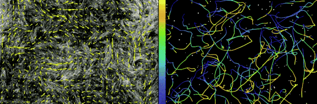
 understood in any organism, not even in simple bacteria.
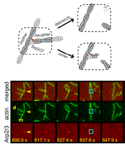 Two separate members of the larger ADF-homology family of proteins, glia maturation factor (GMF) and cofilin, have been implicated in promoting debranching. In this paper, Gelles lab member Johnson Chung, in collaboration with Jeff and with Bruce Goode from the Brandeis Biology Dept., used multi-wavelength single molecule florescence microscopy and quantitative kinetic analysis to define the mechanisms by which these proteins promote debranching. Dr. Chung shows that “cofilin, like GMF, is an authentic debrancher independent of its filament-severing activity and that the debranching activities of the two proteins are additive. While GMF binds directly to the Arp2/3 complex, cofilin selectively accumulates on branch–junction daughter filaments in tropomyosin-decorated networks just prior to debranching events. Quantitative comparison of debranching rates with the known kinetics of cofilin–actin binding suggests that cofilin occupancy of a particular single actin site at the branch junction is sufficient to trigger debranching. In rare cases in which the order of departure could be resolved during GMF- or cofilin-induced debranching, the Arp2/3 complex left the branch junction bound to the pointed end of the daughter filament, suggesting that both GMF and cofilin can work by destabilizing the mother filament–Arp2/3 complex interface. Taken together, these observations suggest that GMF and cofilin promote debranching by distinct yet complementary mechanisms.”
 Congratulations to Dr. Deborah Tenenbaum, shown here with her two Ph.D. mentors Jeff Gelles (smiling under the mask) and
 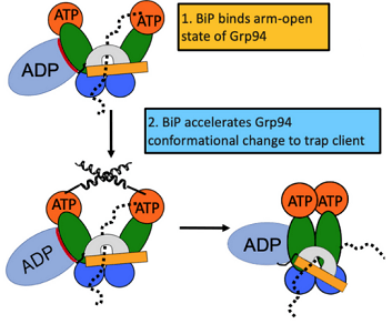 From the article: “The endoplasmic reticulum (ER) is the site at which secreted proteins (such as the hormone insulin) and membrane-bound proteins are folded. ATP-dependent chaperones within the ER help proteins fold. This study describes how two key ER chaperones, BiP and Grp94, work together at a molecular level. BiP binds to Grp94, which enables Grp94 to change conformation and hydrolyze ATP. In short, BiP provides a signal to switch on Grp94 conformational changes that are required to help other proteins fold. This finding helps explain how two chaperones can work together collaboratively in protein folding. Because BiP and Grp94 are members of highly conserved chaperone families, these findings may provide insight into chaperone-assisted protein folding beyond the ER.” This project was a collaboration with members of